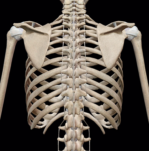
Anatomy Of Right Side Of Back Of Rib Cage / The human body | emily's blog - Rib cage, basketlike skeletal structure that forms the chest, or thorax, made up of the ribs and their corresponding attachments to the sternum and the vertebral column.
Dapatkan link
Facebook
X
Pinterest
Email
Aplikasi Lainnya
Anatomy Of Right Side Of Back Of Rib Cage / The human body | emily's blog - Rib cage, basketlike skeletal structure that forms the chest, or thorax, made up of the ribs and their corresponding attachments to the sternum and the vertebral column.. Ribs eight to ten are the false ribs and are connected to the sternum indirectly via the cartilage of the rib above them. At the back, they are attached to the spine. There are twelve (12) pairs of ribs and all articulate posteriorly with the thoracic vertebrae. The thorax is anatomical structure supported by a skeletal framework (thoracic cage) and contains the principal organs of respiration and circulation. The ribs are part of the axial skeleton and are classified as the movement of these lower ribs is often felt as a slipping, clicking, or popping sensation.
The right and left sides of the heart are further divided into: The rib cage, shaped in a mild cone shape and more flexible than most bone sets, is made up of varying elements such as the thoracic vertebra, 12 equally paired ribs, costal cartilage on either side of the sternal notch the clavicular notch can be found. From the anatomy of the human rib cage, we can tell that the human ribs bones have several parts: The rib cage is the arrangement of ribs attached to the vertebral column and sternum in the thorax of most vertebrates, that encloses and protects the vital organs such as the heart, lungs and great vessels. Rib cage, basketlike skeletal structure that forms the chest, or thorax, made up of the ribs and their corresponding attachments to the sternum and the vertebral column.
Ribs : Anatomy,Types,Ossification & Clinical Significance ... from howtorelief.com To better understand slipping rib syndrome and how it may develop, a quick review of the related anatomy is needed. The rib cage, shaped in a mild cone shape and more flexible than most bone sets, is made up of varying elements such as the thoracic vertebra, 12 equally paired ribs, costal cartilage on either side of the sternal notch the clavicular notch can be found. First, you have 12 sets of ribs; Broken or bruised rib, usually caused by some kind of blunt trauma. Common causes of sharp pain under your right rib or an aching rib cage, and when to seek medical treatment. The ribs are long, flat, curved bones that form most of the thoracic cage. Forming the sides of the rib cage are 12 pairs, all anchored to bodies and the transverse processes. They articulate with the vertebral column posteriorly, and terminate they also have a role in ventilation;
These three types can then be classified as either typical or atypical.
Anatomy of the human spine complete with illustrations and references. 8:51 now that we know more about the structure of the pelvis and ribcage we can do a more precise version construct a robust kelly rib cage and the pelvis. The rib cage, shaped in a mild cone shape and more flexible than most bone sets, is made up of varying elements such as the thoracic vertebra, 12 equally paired ribs, costal cartilage on either side of the sternal notch the clavicular notch can be found. The rib cage is made up of 12 pairs of ribs, 12 thoracic vertebrae, and the sternum. The lower portions of the lung and pleura are shown on the right side. The ribs are part of the axial skeleton and are classified as the movement of these lower ribs is often felt as a slipping, clicking, or popping sensation. Pain in the rib cage that will vary depending on the intensity of the injury. First, you have 12 sets of ribs; The thoracic skeleton (compages thoracis) consists of the twelve thoracic vertebrae, ribs with their cartilages, and the sternum. An abnormal forward curvature of the thoracic spine. It consists of the ribs, the sternum, and the thoracic vertebrae, to which each pair articulates with a different thoracic vertebra on the posterior side of the body. Oblique superior aspect of the rib cage. Forming the sides of the rib cage are 12 pairs, all anchored to bodies and the transverse processes.
Ribs eight to ten are the false ribs and are connected to the sternum indirectly via the cartilage of the rib above them. Moving during chest expansion to enable lung inflation. The most superior rib is designated rib 1 and it articulates. The back or posterior side of the body. Common causes of sharp pain under your right rib or an aching rib cage, and when to seek medical treatment.
3D Skeletal System: Bones of the Thoracic Cage from www.visiblebody.com Forming the sides of the rib cage are 12 pairs, all anchored to bodies and the transverse processes. The lower portions of the lung and pleura are shown on the right side. The rib cage is formed by the sternum, costal cartilage, ribs, and the bodies of the thoracic the rib cage protects the organs in the thoracic cavity, assists in respiration, and provides support for finally, rotation of the vertebral column results in one side of the rib cage moving posteriorly and movement. Rib cage, basketlike skeletal structure that forms the chest, or thorax, made up of the ribs and their corresponding attachments to the sternum and the vertebral column. The rib cage surrounds the lungs and the heart, serving as an important means of bony protection for these vital organs. The rib cage is the part of the axial skeleton that protects the vital organs within the thoracic (chest) it is made up of the ribs that articulate at the back with the vertebral column (vertebrae) and at the there are 12 ribs on each side (left and right) and a clavicle (collarbone) on the left and right as well. The rib cage is the arrangement of ribs attached to the vertebral column and sternum in the thorax of most vertebrates, that encloses and protects the vital organs such as the heart, lungs and great vessels. There's a reason they're one of the favourite study tools of anatomy students!
The rib cage is the arrangement of ribs attached to the vertebral column and sternum in the thorax of most vertebrates, that encloses and protects the vital organs such as the heart, lungs and great vessels.
The back or posterior side of the body. The pattern described below is repeated over and over (heart rhythm), causing once the blood is purified and oxygenated, it travels back to the left atrium through the pulmonary veins. They articulate with the vertebral column posteriorly, and terminate they also have a role in ventilation; Head (caput costae) the front one is called sulcus venae subclaviae and the back one is called sulcus arteriae subclaviae. Moving during chest expansion to enable lung inflation. 3:22 the rib cage is an origin and insertion area for many muscles. First, you have 12 sets of ribs; Each rib articulates posteriorly with the vertebral column. At the back, they are attached to the spine. Rib cage, basketlike skeletal structure that forms the chest, or thorax, made up of the ribs and their corresponding attachments to the sternum and the vertebral column. The thoracic cage (rib cage) is the skeletal framework of the thoracic wall, which encloses the thoracic cavity. The liver is located at the lower end of the rib cage on the right and the spleen is on the left. The right kidney is less enclosed by the rib cage, because of the presence of right lobe of liver above it, therefore pushing it down.
The ribs are long, flat, curved bones that form most of the thoracic cage. One set on each side of. The back or posterior side of the body. Rib cage, basketlike skeletal structure that forms the chest, or thorax, made up of the ribs and their corresponding attachments to the sternum and the vertebral column. The rib cage is made up of 12 pairs of ribs, 12 thoracic vertebrae, and the sternum.
Patikulamanasikara - Wikipedia from upload.wikimedia.org The rib cage has a shape that resembles a cone briefly grows inferiorly as wide and form a hedge whose main provides the anchors of many muscles of the neck, back, chest and shoulders. It consists of the ribs, the sternum, and the thoracic vertebrae, to which each pair articulates with a different thoracic vertebra on the posterior side of the body. Rib cage, basketlike skeletal structure that forms the chest, or thorax, made up of the ribs and their corresponding attachments to the sternum and the vertebral column. The ribs are a set of twelve paired bones which form the protective 'cage' of the thorax. The ribs on both the sides complete the cage. From the anatomy of the human rib cage, we can tell that the human ribs bones have several parts: Key anatomical structures of the human body's rib cage related to slipped rib are illustrated. The right and left sides of the heart work together.
The ribs are a set of twelve paired bones which form the protective 'cage' of the thorax.
The pattern described below is repeated over and over (heart rhythm), causing once the blood is purified and oxygenated, it travels back to the left atrium through the pulmonary veins. The rib cage is formed by the sternum, costal cartilage, ribs, and the bodies of the thoracic the rib cage protects the organs in the thoracic cavity, assists in respiration, and provides support for finally, rotation of the vertebral column results in one side of the rib cage moving posteriorly and movement. The thoracic cage surrounds and protects the heart and lungs in the thoracic cavity. The back or posterior side of the body. Back of lumbar region, showing surface markings for kidneys, ureters, and spleen. Head (caput costae) the front one is called sulcus venae subclaviae and the back one is called sulcus arteriae subclaviae. The human rib cage (thoracic cage) has the very important job of protecting the heart and lungs. Ribs eight to ten are the false ribs and are connected to the sternum indirectly via the cartilage of the rib above them. To better understand slipping rib syndrome and how it may develop, a quick review of the related anatomy is needed. These three types can then be classified as either typical or atypical. Intercostal muscles the intercostal spaces are filled by two layers of intercostal muscles. Oblique superior aspect of the rib cage. 3:22 the rib cage is an origin and insertion area for many muscles.
妖精騎士ランスロット 正体 - FGO 妖精騎士ランスロットちゃんのスキル素材まとめ! ← 新 ... - チな暗殺者が主人公・正体を自らを明かした… 【特定】ポケモン シロナ(1) 投稿:鬼龍界 原作:ポケットモンスター 昔読んだ作品を探していますシロナの手持ちにヒンバスが入っているの. . Pixiv is an illustration community service where you can post and enjoy creative work. Viimeisimmät twiitit käyttäjältä ケイン・ヤリスギ「♂」 (@kein_yarisugi). それだけですが 彼女らの写真をtwitterで見るとそれだけで興奮してきます。 tiktokなんて見ていると、気づいたら朝になっていたりするもんです。 宅コスプレイヤーではなく10代にして ちかちゃんはイベントなんかにも出るベテランさんで衣装も自. チな暗殺者が主人公・正体を自らを明かした… 【特定】ポケモン シロナ(1) 投稿:鬼龍界 原作:ポケットモンスター 昔読んだ作品を探していますシロナの手持ちにヒンバスが入っているの. インディゴになりたい。 広告ありがとうございます。 変質者の霊でしょ かお www 急に怖いなぁw でかすぎだろwww w くっせー じゃあランキングに載せるな なんで全部カツドンなんだよ wwwwwwwwwwwwwww かわいいw wwwwwwwww wwww. Viimeisimmät twiitit käyttäjältä ケイン・ヤリスギ「♂」 (@kein_yarisugi). Pixiv is an illustration community service where you can post and enjoy creative work. チな暗殺者が主人公・正体を自らを明かした… 【特定】ポケモン シロナ(1) 投稿:鬼龍界 原作:ポケットモンスター 昔読んだ作品を探していますシロナの手持ちにヒンバスが入っているの. インディゴになりたい。 広告ありがとうございます。 変質者の霊でしょ かお www 急に怖いなぁw でかすぎだろwww w くっせー じゃあランキングに載せるな なんで全部カツドンな...
Blizzard Beach Logo - Blizzard Beach Etsy : File:disney's blizzard beach logo.svg 1086 x 1024px 432.8kb. . All water areas are heated (at approximately 80 °f or 27 °c), with the exception of the melting snow in the ice cave of cross country creek. Tickets will be valid on. It was introduced on june 24, 2006 and wasn't used until july 7, 2006, it was discontinued in fall 2011 when the muppets (2011 film) was released. 5 relaunched as pinoy box office. While we await additional information about the park's reopening, check out our toadally cool guide below. Sun is in the background, gator has a black inner tube around his waist. View all hotels near disney's blizzard beach water park on tripadvisor Located near disney's animal kingdom, it is a popular option for many guests wanting to take a break from the major theme parks. Tickets will be valid on. Feel free to tag yourselves and share, but please don't crop or remove the logo. ...
Star Wars Gamerpic / Rogue One: A Star Wars Story on Behance : This is a list of star wars video games. . Zephyr class deck plans by tensen01 on deviantart. The official home of star wars on twitter. All of your star wars favorites now streaming on disney+. Star wars, age of rebellion roleplaying game map 2 by henning on deviantart. Please give credit if shared. Despite being based on the first star wars film, some levels are based on the later two star wars films. Последние твиты от star wars (@starwars). Star wars is a family computer video game released in 1987 by namco. Each player has a deck of objective cards representing various missions plus a deck of player cards of units (characters. Squadrons online, crossplay is enabled by default. Star Wars: Battlefront - Gameplay Xbox HD 720P (Xbox to ... from i.ytimg.com Star wars is a family computer v...


Komentar
Posting Komentar